TRAUMA YELLOW
  • Clinical
    • Discharge Macros
    • Procedure Macros
    • Exam Macros
    • Pediatric Macros
    • Antibiogram
    • Follow Up
    • 2023 MDM
    • Heart Pathway
    • Jeromy's Macros
  • Education
    • EM Education
    • Critical Care
    • FOAM
  • Orientation
    • Dept Orientation
    • Cerner
    • Dragon
  • Links
    • Clinical Links
    • StatMacros
    • ICU Bootcamp
  • Private
    • MCS
    • GWU Contacts & Map

Management of Deep Vein Thrombosis

2/7/2018

0 Comments

 
In the US, approximately 200,000 people are diagnosed with a new or recurrent pulmonary embolism each year. More than twice this many are diagnosed with a DVT without PE, and 1 in 300 ER patients are diagnosed with a DVT. The lifetime risk of a DVT is 2-5%, and the incidence of DVT increases with age. DVTs can lead to a pulmonary embolism, which is one of the leading causes of sudden, nontraumatic death in the US. 

Most DVTs are unprovoked, with an increased risk for reoccurrence, requiring a longer treatment period than those with provoked DVTs from drugs, trauma, or surgery.
Typically, DVTs begin at the valves of the veins. Proximal DVT include Femoropopliteal (FP) ilieofemoral (IF) DVTs, and account for 80% of symptomatic DVTs. Distal DVTs of the calf account for only 20% of symptomatic DVTs. 

Diagnosis

A DVT typically presents with unilateral pain and swelling of a lower extremity, but patients may also complain of cramping or pain with use. Lower DVTs are more likely to be associated with thrombophlebitis. An upper-extremity may present similarly to cellulitis, and those with long-term catheters, such as PICC lines, are at increased risk. Finger swelling or cellulitic-like changes may suggest an upper-extremity DVT. 
  • The calves should be measured 10cm below the tibial tubercle. A difference >2cm between leg diameter is concerning for a DVT. 
  • Pain elicited with passive dorsiflexion of the ankle, called Homan's Sign, is neither sensitive nor specific for an upper or lower DVT. 
Patients concerning for a DVT should have a cardiopulmonary evaluation, as well as a duplex ultrasound to evaluate for DVT. Patients at low-risk should first be evaluated with the Well's Criteria for DVT, and may instead have a d dimer instead of US testing. An ultrasound can have sensitivity up to 90%, and 95% specificity to diagnosis DVT. 

Patients with a positive d dimer and negative US should have repeat US testing within the next week. 

Treatment

Patients with DVT require systemic anticoagulation, with either heparin or low molecular weight heparin, while rivaroxaban can be used in many cases, it is not approved for DVTs in the setting of cancer. Those with renal disease should be put on heparin or LMWH. 

Treatment of isolated calf thrombosis has no set guidelines; some recommend no treatment with followup imaging, while others recommend outpatient LMWH. 

Indications for Admission of DVT
  • An extensive iliofemoral deep venous thrombosis with circulatory compromise
  • An increased risk of bleeding (coagulopathy, active peptic ulcer disease, liver disease) that requires close monitoring of therapy
  • A limited cardiorespiratory reserve
  • A risk of poor compliance or poor followup
  • Known or suspected PE that is high-risk
  • History of Heparin-induced thrombocytopenia
  • Renal insufficiency

Complications

Post-thrombotic Syndrome (PTS) occurs in 25-50% of patients with proximal DVTs and develops over months to years after a DVT, manifesting with swelling, erythema, and pain. This is due to the scaring and sclerosis of valves, leading to poor function. Skin thickening and recurrent DVTs may occur. Compression stocks can reduce the incidence, however there is low compliance with therapy. 

Phlegmasia Cerulea Dolens: A rare complete, extensive venous occlusion of the leg, with severe pain, edema, and cyanosis. Despite intervention, there is a high risk of PE, as well as gangrene of the foot. 

May Thurner Syndrome: Left iliac vein compression by the iliac artery, leading to large femoral DVTs and requiring surgical intervention and endovascular catheter-directed lysis, aspiration, and angioplasty. 
Picture
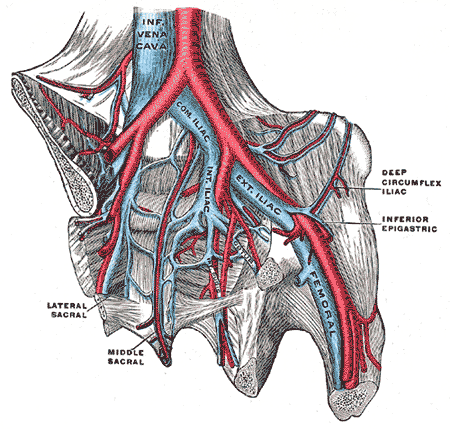
https://commons.wikimedia.org/wiki/File:Iliac_veins.gif
0 Comments


Leave a Reply.

    Categories


    All
    Abx
    Article
    Cardiac
    EKG
    EKG Challenge
    GI Bleed
    Grand Rounds
    Headache
    ICU
    Orthopedic
    Pain
    Peds
    Reading
    Stroke
    Subarachnoid Hemorrhage
    Syncope
    Teaching Pearl
    Trauma
    Travel
    Zika

    Archive


    February 2018
    January 2018
    December 2017
    November 2017
    October 2017
    September 2017
    August 2017
    May 2017
    April 2017
    March 2017
    February 2017
    January 2017
    December 2016
    November 2016
    October 2016
    September 2016
    August 2016
    July 2016
    June 2016
    May 2016
    April 2016
    January 2016
    December 2015
    November 2015

    Picture
    Please read our Terms of Use.
Donate
This site is independently owned and operated and not affiliated with the George Washington University, George Washington University Hospital, or Medical Faculty Associates. All information on this site is the opinion of the author alone and in no way should be seen to represent the views of the George Washington University, George Washington University Hospital, or Medical Faculty Associates. The information on this page is for personal use only and should not be see as medical advice or used directly for patient care. The author provides no guarantee of the accuracy of the information provided on this page. ​

​Please refer to our Terms of Use. 
  • Clinical
    • Discharge Macros
    • Procedure Macros
    • Exam Macros
    • Pediatric Macros
    • Antibiogram
    • Follow Up
    • 2023 MDM
    • Heart Pathway
    • Jeromy's Macros
  • Education
    • EM Education
    • Critical Care
    • FOAM
  • Orientation
    • Dept Orientation
    • Cerner
    • Dragon
  • Links
    • Clinical Links
    • StatMacros
    • ICU Bootcamp
  • Private
    • MCS
    • GWU Contacts & Map