TRAUMA YELLOW
  • Clinical
    • Discharge Macros
    • Procedure Macros
    • Exam Macros
    • Pediatric Macros
    • Antibiogram
    • Follow Up
    • 2023 MDM
    • Heart Pathway
    • Jeromy's Macros
  • Education
    • EM Education
    • Critical Care
    • FOAM
  • Orientation
    • Dept Orientation
    • Cerner
    • Dragon
  • Links
    • Clinical Links
    • StatMacros
    • ICU Bootcamp
  • Private
    • MCS
    • GWU Contacts & Map

Lower GI Bleeding

8/16/2017

0 Comments

 
Picture
Adapted from Katie Newell's August 16th 2017 Grand Rounds Lecture. 
Lower GI bleeding refers to any bleeding below the Ligament of Treitz and can be due to many causes, some highly dangerous and others less so. Females are more likely to have a lower GI bleed, and the risk increases significantly in elderly patients. 80% of lower GI bleeds resolve spontaneously, however it is difficult to assess prior to determining the exact cause of the bleed.

Causes of Lower GI Bleeding

Colorectal sources, such as hemorrhoids, are the most common causes of LGIB in all age groups, followed by diverticular disease, colitis, polyps, and maligancies. Diverticular bleeding from an eroded artery is painless and can be brisk, but typically is self resolving, but frequently recur. Right-sided diverticulosis is more likely to bleed, although left-sided diverticulosis is more common. A concomitant diverticulitis is exceptionally rare with a bleeding diverticula. 

Vascular causes, such as an AV malformation or angiodysplasia can occur in the small or large bowel, and typically associated with a chronic process and increases with aging. These are less commonly occurring. 

Meckel's diverticulum contain ectopic gastric tissue, which can secrete gastric enzymes, eroding the mucosal wall and causing bleeding. Meckel's is rare, but can be an important cause of LGI bleeding in younger patients. 

There are numerous other causes for LGI bleeding, including infectious colitis, inflammatory bowel disease, trauma, or anticoagulation use. 

Determining the Source

Hematochezia is bright red or maroon-colored rectal bleeding, which indicates either a brisk UGI or a lower GI bleed. Hematemesis and hemodynamic instability should accompany a brisk UGI bleed. Melena is dark or black-colored stools which typically represents an UGI bleed, but could represent a LGI source. ​​

Imaging

Picture
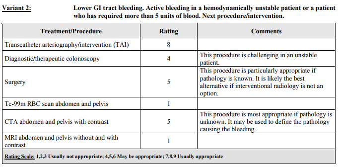
ACR recommends CTA in those patients who have an unknown source of LGI bleeding, and may be used to define the pathology causing the bleeding. The tagged RBC scan has fallen out of use in unstable patients with an ACR rating of 1. 

Disposition

High-risk populations include:
  • Age >60
  • Oral corticosteroids
  • Diabetes mellitus, COPD, Current smoking, NSAID use,
  • Long-term aspirin use 
  • Syncope or near-syncope as presenting complaint
  • Elevated BUN, prothrombin time or other lab abnormalities
  • Those to be admitted to the ICU include those with current hemodynamic instability and those with the potential to decompensate.

Low-risk population include;
  • No “severe anemia” (Hgb <8 g/dL)
  • Age <60
  • Stable Vital Signs
  • Pulse < 100 bpm
  • No fresh, voluminous hematemesis or multiple complains of melena on the day of presentation
  • No concomitant disease*
  • No anticoagulation
​
​Currently, there is no reliable scoring system to risk stratify patients with LGI bleeding. Patients may be candidates for discharge with followup are those who have a mild LGI bleed with an obvious source (such as hemorrhoids), who have no bright red blood or maroon/melanotic stool on examination, and are hemodynamically stable without major comorbidities. 
​
0 Comments


Leave a Reply.

    Categories


    All
    Abx
    Article
    Cardiac
    EKG
    EKG Challenge
    GI Bleed
    Grand Rounds
    Headache
    ICU
    Orthopedic
    Pain
    Peds
    Reading
    Stroke
    Subarachnoid Hemorrhage
    Syncope
    Teaching Pearl
    Trauma
    Travel
    Zika

    Archive


    February 2018
    January 2018
    December 2017
    November 2017
    October 2017
    September 2017
    August 2017
    May 2017
    April 2017
    March 2017
    February 2017
    January 2017
    December 2016
    November 2016
    October 2016
    September 2016
    August 2016
    July 2016
    June 2016
    May 2016
    April 2016
    January 2016
    December 2015
    November 2015

    Picture
    Please read our Terms of Use.
Donate
This site is independently owned and operated and not affiliated with the George Washington University, George Washington University Hospital, or Medical Faculty Associates. All information on this site is the opinion of the author alone and in no way should be seen to represent the views of the George Washington University, George Washington University Hospital, or Medical Faculty Associates. The information on this page is for personal use only and should not be see as medical advice or used directly for patient care. The author provides no guarantee of the accuracy of the information provided on this page. ​

​Please refer to our Terms of Use. 
  • Clinical
    • Discharge Macros
    • Procedure Macros
    • Exam Macros
    • Pediatric Macros
    • Antibiogram
    • Follow Up
    • 2023 MDM
    • Heart Pathway
    • Jeromy's Macros
  • Education
    • EM Education
    • Critical Care
    • FOAM
  • Orientation
    • Dept Orientation
    • Cerner
    • Dragon
  • Links
    • Clinical Links
    • StatMacros
    • ICU Bootcamp
  • Private
    • MCS
    • GWU Contacts & Map