TRAUMA YELLOW
  • Clinical
    • Discharge Macros
    • Procedure Macros
    • Exam Macros
    • Pediatric Macros
    • Antibiogram
    • Follow Up
    • 2023 MDM
    • Heart Pathway
    • Jeromy's Macros
  • Education
    • EM Education
    • Critical Care
    • FOAM
  • Orientation
    • Dept Orientation
    • Cerner
    • Dragon
  • Links
    • Clinical Links
    • StatMacros
    • ICU Bootcamp
  • Private
    • MCS
    • GWU Contacts & Map

Diagnosis and Management of Pancreatitis: an evidence based approach

8/30/2017

0 Comments

 
Adapted from Katie Januszewicz's Grand Rounds lecture August 30th, 2017
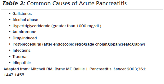
Inflammation of the pancreas can be limited to mild inflammation to severe necrosis of the pancreas and surrounding tissue. Most cases (80%) involve only mild inflammation, however severe pancreatitis can lead to a 30% mortality rate. Most cases are secondary to gallstones or alcohol, but up to 5% of ERCP patients present with post-procedure pancreatitis. Additionally, many medications (120) can cause pancreatitis. In severe cases this can lead to inflammatory responses in nearby tissue and even systemically.  ​

Pancreatitis occurs secondary to an unclear mechanism involving trypsin, a protease that is typically converted from pancreatic trypsinogen in the duodenum. When trypsin is activated in the pancreatic acinar cells, this continues unregulated leading to activation of other digestive enzymes and auto-digestion of the pancreas. 
​
Picture

Diagnosis

Diagnosis is based on at least two of three criteria;
  • Clinical presentation: Epigastric, radiating abdominal pain
  • Elevated Serum Lipase or amylase more than 3x normal upper limit
  • Imaging findings characteristic of acute pancreatitis on CT with IV contrast
Current guidelines recommend that amylase or lipase should be elevated at least three times normal, although some guidelines recommend any lipase elevation is abnormal. Overall, amylase is 70% sensitive and a positive-predictive-value  of 15-72%. Amylase can be elevated in multiple non-pancreas diseases such as appendicitis, renal insufficiency, and intestinal obstruction. Lipase is more specific and remains elevated longer after onset of symptoms. 

ALT elevation > 150 U/L within the first 48 hours of symptoms has a 85% positive predictive value of gallstone pancreatitis. 

Reserve CT imaging for cases of diagnostic uncertainty, severe pancreatitis, especially if high suspicion of complications (pseudocyst, necrosis). Abdominal ultrasound can be helpful in confirming cases of gallstone pancreatitis, but is not the gold standard for pancreatitis imaging.

​There is no evidence that early CT improves clinical outcomes. 
​
yield_of_early_ct_scan_in_pancreatitis.pdf
File Size: 129 kb
File Type: pdf
Download File

Management & Disposition

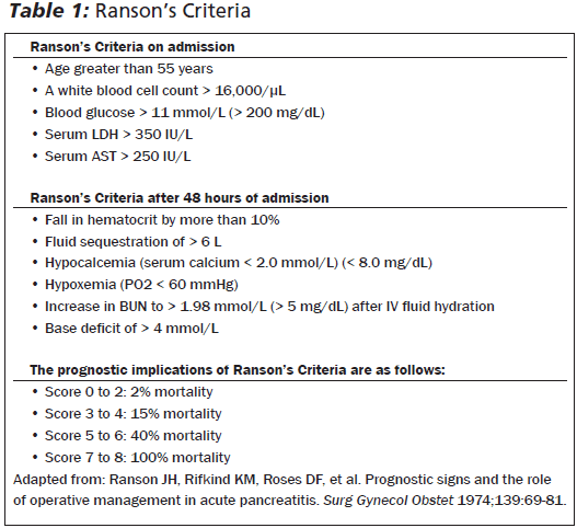
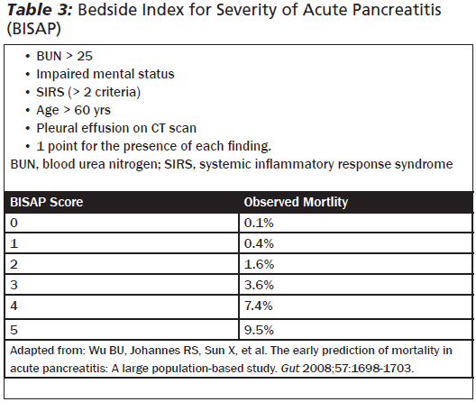
Mainstay of management is supportive care (analgesia, antiemetics, maintenance IV fluids). Diet should be NPO, unless the case is mild and the patient can tolerate PO. Prophylactic antibiotics are not recommended. Scoring systems can be helpful to determine mild to severe cases of pancreatitis. In the ED, it is easiest to obtain a BISAP score for mortality risk (as compared to a Ranson score which is becoming outdated). Disposition ranges from discharge (mild case, tolerating PO) to ICU admissions (hemodynamically unstable, severe pancreatitis, signs of end organ damage). The rest can be admitted to the floor and may require surgical consults (if planning cholecystectomy, etc.) or GI consults (for ERCP).
Picture
Picture
0 Comments


Leave a Reply.

    Categories


    All
    Abx
    Article
    Cardiac
    EKG
    EKG Challenge
    GI Bleed
    Grand Rounds
    Headache
    ICU
    Orthopedic
    Pain
    Peds
    Reading
    Stroke
    Subarachnoid Hemorrhage
    Syncope
    Teaching Pearl
    Trauma
    Travel
    Zika

    Archive


    February 2018
    January 2018
    December 2017
    November 2017
    October 2017
    September 2017
    August 2017
    May 2017
    April 2017
    March 2017
    February 2017
    January 2017
    December 2016
    November 2016
    October 2016
    September 2016
    August 2016
    July 2016
    June 2016
    May 2016
    April 2016
    January 2016
    December 2015
    November 2015

    Picture
    Please read our Terms of Use.
Donate
This site is independently owned and operated and not affiliated with the George Washington University, George Washington University Hospital, or Medical Faculty Associates. All information on this site is the opinion of the author alone and in no way should be seen to represent the views of the George Washington University, George Washington University Hospital, or Medical Faculty Associates. The information on this page is for personal use only and should not be see as medical advice or used directly for patient care. The author provides no guarantee of the accuracy of the information provided on this page. ​

​Please refer to our Terms of Use. 
  • Clinical
    • Discharge Macros
    • Procedure Macros
    • Exam Macros
    • Pediatric Macros
    • Antibiogram
    • Follow Up
    • 2023 MDM
    • Heart Pathway
    • Jeromy's Macros
  • Education
    • EM Education
    • Critical Care
    • FOAM
  • Orientation
    • Dept Orientation
    • Cerner
    • Dragon
  • Links
    • Clinical Links
    • StatMacros
    • ICU Bootcamp
  • Private
    • MCS
    • GWU Contacts & Map