TRAUMA YELLOW
  • Clinical
    • Discharge Macros
    • Procedure Macros
    • Exam Macros
    • Pediatric Macros
    • Antibiogram
    • Follow Up
    • 2023 MDM
    • Heart Pathway
    • Jeromy's Macros
  • Education
    • EM Education
    • Critical Care
    • FOAM
  • Orientation
    • Dept Orientation
    • Cerner
    • Dragon
  • Links
    • Clinical Links
    • StatMacros
    • ICU Bootcamp
  • Private
    • MCS
    • GWU Contacts & Map

Describing Fractures

11/30/2016

0 Comments

 
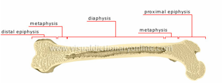
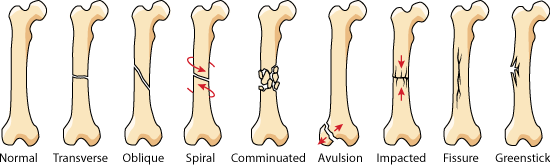
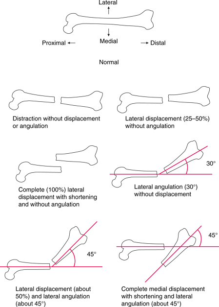
When consulting for fracture care, it is important to provide an accurate description of findings over the phone. This is important to improve patient outcome, ensure consultants are well informed of the patient's condition, and to ensure appropriate documentation of a patient's injuries.
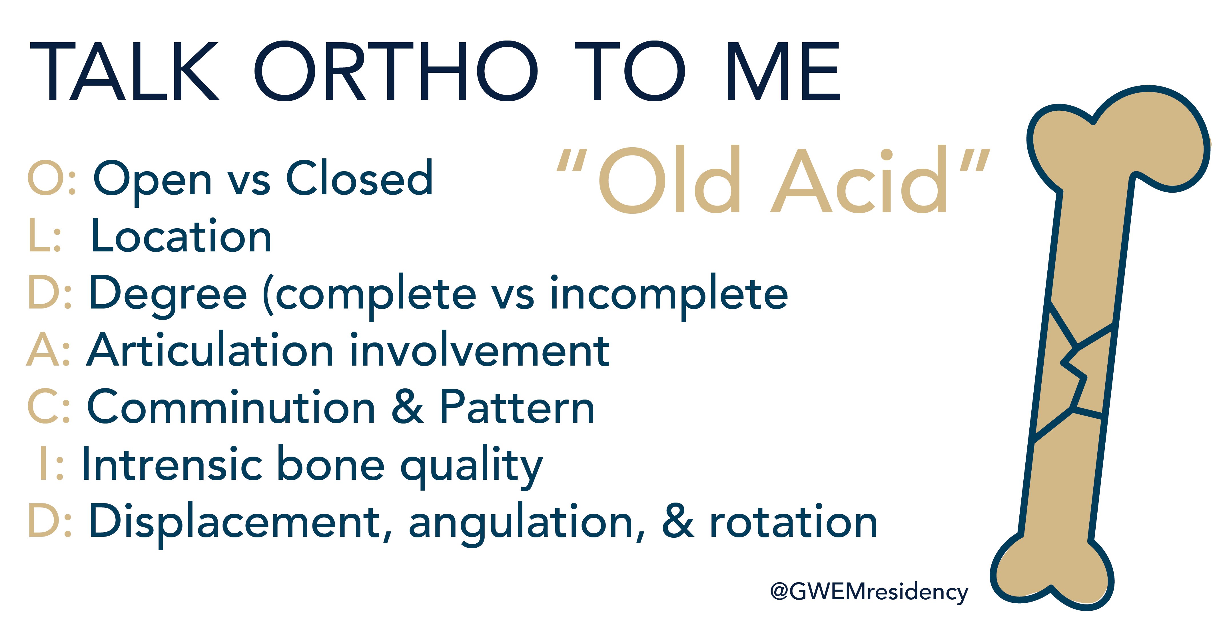
Picture

Wet read

Every potential fracture site requires at least two views to fully evaluate for injury. You should also be imaging the joint above and below the injury as well. When reading your imaging, make sure you identify any joint involvement, and second fractures that may be present, as well as lines and tubes. 

Naming fractures

The menemonic OLD ACID can be used to describe fractures
  • O: Open vs Closed Is the fracture open, or 'compound' i.e. open to air 
  • L: Location Which bone is involved, and what part of the bone? In long bones, identify the epiphysis, metaphysis, diaphysis, or physis, as well as the articular surface. Anatomic landmarks, such as head, neck, body, shaft, base, or condyle. 
Picture
  • D: Degree of fracture A complete fracture will include the entire cortical circumference, and fragments are completely separated. Incomplete fractures, such as a Greenstick fracture, does not compromise the entire cortex.
  • A: Articular Extension Identify any involvement of the intra-articular surface, as well as any dislocation.
  • C: Comminution/ Pattern Fractures can have multiple patterns of fracture, depending on the forces involved. Atypical fracture patterns include impacted, hairline, torus (buckle) and pathologic fractures. Bones with multiple fractures are segmental, and should be called comminuted if there are three or more segments.
Picture
  • I: Intrinsic bone quality Identify any osteopetrosis or osteopenia
  • D: Displacement, angulation, rotation. Displacement idenfities fracture fragments are not axially aligned. Identify displacement of distal compared to medial. i.e. laterally displaced. Angulation of a fracture is relative to the anatomic position of the fracture compared to the proximal bone. Medial angulation is Varus Angulation, while lateral angulation is Valgus Angulation. Rotation of a fracture is either internal or external. 
Picture

Specific consult question

When consulting, ensure you have a specific consult question, especially when a consultant may not be in the hospital or may not have access directly to imaging. 
0 Comments


Leave a Reply.

    Categories


    All
    Abx
    Article
    Cardiac
    EKG
    EKG Challenge
    GI Bleed
    Grand Rounds
    Headache
    ICU
    Orthopedic
    Pain
    Peds
    Reading
    Stroke
    Subarachnoid Hemorrhage
    Syncope
    Teaching Pearl
    Trauma
    Travel
    Zika

    Archive


    February 2018
    January 2018
    December 2017
    November 2017
    October 2017
    September 2017
    August 2017
    May 2017
    April 2017
    March 2017
    February 2017
    January 2017
    December 2016
    November 2016
    October 2016
    September 2016
    August 2016
    July 2016
    June 2016
    May 2016
    April 2016
    January 2016
    December 2015
    November 2015

    Picture
    Please read our Terms of Use.
Donate
This site is independently owned and operated and not affiliated with the George Washington University, George Washington University Hospital, or Medical Faculty Associates. All information on this site is the opinion of the author alone and in no way should be seen to represent the views of the George Washington University, George Washington University Hospital, or Medical Faculty Associates. The information on this page is for personal use only and should not be see as medical advice or used directly for patient care. The author provides no guarantee of the accuracy of the information provided on this page. ​

​Please refer to our Terms of Use. 
  • Clinical
    • Discharge Macros
    • Procedure Macros
    • Exam Macros
    • Pediatric Macros
    • Antibiogram
    • Follow Up
    • 2023 MDM
    • Heart Pathway
    • Jeromy's Macros
  • Education
    • EM Education
    • Critical Care
    • FOAM
  • Orientation
    • Dept Orientation
    • Cerner
    • Dragon
  • Links
    • Clinical Links
    • StatMacros
    • ICU Bootcamp
  • Private
    • MCS
    • GWU Contacts & Map