TRAUMA YELLOW
  • Clinical
    • Discharge Macros
    • Procedure Macros
    • Exam Macros
    • Pediatric Macros
    • Antibiogram
    • Follow Up
    • 2023 MDM
    • Heart Pathway
    • Jeromy's Macros
  • Education
    • EM Education
    • Critical Care
    • FOAM
  • Orientation
    • Dept Orientation
    • Cerner
    • Dragon
  • Links
    • Clinical Links
    • StatMacros
    • ICU Bootcamp
  • Private
    • MCS
    • GWU Contacts & Map

Blunt Thoracic Trauma

10/19/2016

0 Comments

 
Adapted from Kristin Schreiber's Grand Rounds presentation, October 18th, 2016
Picture

Blunt Cardiac Injury

Any traumatic injury which effects the heart's ability to function. Normally diagnosed via EKG and blood monitoring. Commonly caused by direct trauma to the chest, including MVC, pedestrians, and crush injuries. Injuries can range from pericardial injury or cardiac rupture, to mild troponin elevations and sinus tachycardia. 

Patients at risk for cardiac injury
  • Seatbelt sign
  • Ecchymosis or tenderness to the chest wall 
  • Sternal fractures or rib fractures
  • Subcutaneous air
  • Severe mechanism of injury

Treatment Guidelines

East Trauma Guidelines, 2012
Level 1 Guidelines: 
  • Obtain an EKG for any patient suspected of BCI
Level 2 Guidelines:
  • Admission for any new EKG changes other than sinus tachycardia
  • Elevated troponins, regardless of EKG, should be admitted for monitoring
  • Normal EKGs with normal troponins can be ruled out for BCI, even in patients with sternal fractures
  • Any admission with BCI should have an echocardiogram prior to discharge
  • East Trauma Guidlines recommended against CK utilization
screening_for_blunt_cardiac_injury___an_eastern.5.pdf
File Size: 247 kb
File Type: pdf
Download File

Higher mortality is noted in patients who are hypotensive on presentation, lactate >2.5, co-morbid hemothorax, or elevated troponins. 

Commotio Cordis

Blunt force trauma to the pericordium during the t-wave, causing a fine ventricular fibrillation

Disposition

Patients without other injuries, who are hemodynamically stable with isolated abnormal EKGs or elevated troponins should be admitted for 24-48 hours. 
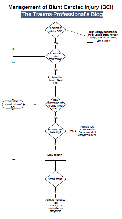
Picture
0 Comments


Leave a Reply.

    Categories


    All
    Abx
    Article
    Cardiac
    EKG
    EKG Challenge
    GI Bleed
    Grand Rounds
    Headache
    ICU
    Orthopedic
    Pain
    Peds
    Reading
    Stroke
    Subarachnoid Hemorrhage
    Syncope
    Teaching Pearl
    Trauma
    Travel
    Zika

    Archive


    February 2018
    January 2018
    December 2017
    November 2017
    October 2017
    September 2017
    August 2017
    May 2017
    April 2017
    March 2017
    February 2017
    January 2017
    December 2016
    November 2016
    October 2016
    September 2016
    August 2016
    July 2016
    June 2016
    May 2016
    April 2016
    January 2016
    December 2015
    November 2015

    Picture
    Please read our Terms of Use.
Donate
This site is independently owned and operated and not affiliated with the George Washington University, George Washington University Hospital, or Medical Faculty Associates. All information on this site is the opinion of the author alone and in no way should be seen to represent the views of the George Washington University, George Washington University Hospital, or Medical Faculty Associates. The information on this page is for personal use only and should not be see as medical advice or used directly for patient care. The author provides no guarantee of the accuracy of the information provided on this page. ​

​Please refer to our Terms of Use. 
  • Clinical
    • Discharge Macros
    • Procedure Macros
    • Exam Macros
    • Pediatric Macros
    • Antibiogram
    • Follow Up
    • 2023 MDM
    • Heart Pathway
    • Jeromy's Macros
  • Education
    • EM Education
    • Critical Care
    • FOAM
  • Orientation
    • Dept Orientation
    • Cerner
    • Dragon
  • Links
    • Clinical Links
    • StatMacros
    • ICU Bootcamp
  • Private
    • MCS
    • GWU Contacts & Map